HEAL-NEURO-CS01-76M-202009
Apparent Reversal of Cerebral Atrophy Following HEAL Method™ Cranial Intervention: A Comparative MRI Case Study
Published/Copyright: 12 August 2025
Abstract:
We present sequential axial brain MRI images of a male patient (born in 1949) obtained during dementia screening in February 2019 and December 2020. Between September and November 2020, the patient underwent five sessions of the HEAL Method™ cranial intervention at the HEAL Institute®.
1. Introduction
Cerebral atrophy is a hallmark neuroimaging finding in various neurodegenerative conditions, including dementia, and is typically considered a progressive and largely irreversible process. Emerging complementary and integrative therapies aim to improve neurological function by influencing cranial biomechanics, cerebrospinal fluid (CSF) dynamics, and neurovascular flow. The HEAL Method™ manual cranial intervention was developed to optimize cranial motion and potentially enhance intracranial physiology. This case report presents sequential MRI findings in a patient with prior evidence of cerebral atrophy before and after a course of HEAL Method™ cranial treatments.
2. Case Presentation
A male patient, born in 1949, underwent routine dementia screening MRI on February 22, 2019. Imaging demonstrated generalized cerebral volume loss, consistent with his clinical profile of cognitive decline. Between September and November 2020, the patient received five sessions of the HEAL Method™ cranial intervention, administered by Dr. Daniel Foo. A follow-up MRI was performed on December 31, 2020, as part of ongoing clinical evaluation.
3. Intervention
The HEAL Method™ cranial intervention consists of a series of manual techniques designed to restore optimal cranial mobility, enhance CSF circulation, and normalize dural membrane tension. Treatments were administered over a three-month period in a private clinical setting. Each session lasted approximately 90 minutes and was tailored to the patient’s cranial biomechanics and clinical presentation. No adverse events were reported.
4. Imaging Findings
February 2019 MRI:
- Prominent cortical sulci
- Widened extra-axial cerebrospinal fluid (CSF) spaces
- Increased distance between the cortical surface and the inner table of the skull
- Cerebellum appearing reduced in relative volume, with enlarged pericerebellar CSF spaces
December 2020 MRI:
- Reduction in extra-axial CSF spaces
- Less prominent cortical sulci
- Brain parenchyma more closely apposed to the calvarium
- Cerebellum more fully occupying the posterior fossa, with decreased pericerebellar CSF volume
These qualitative observations suggest an apparent increase in brain volume or a reduction in extra-axial CSF space.
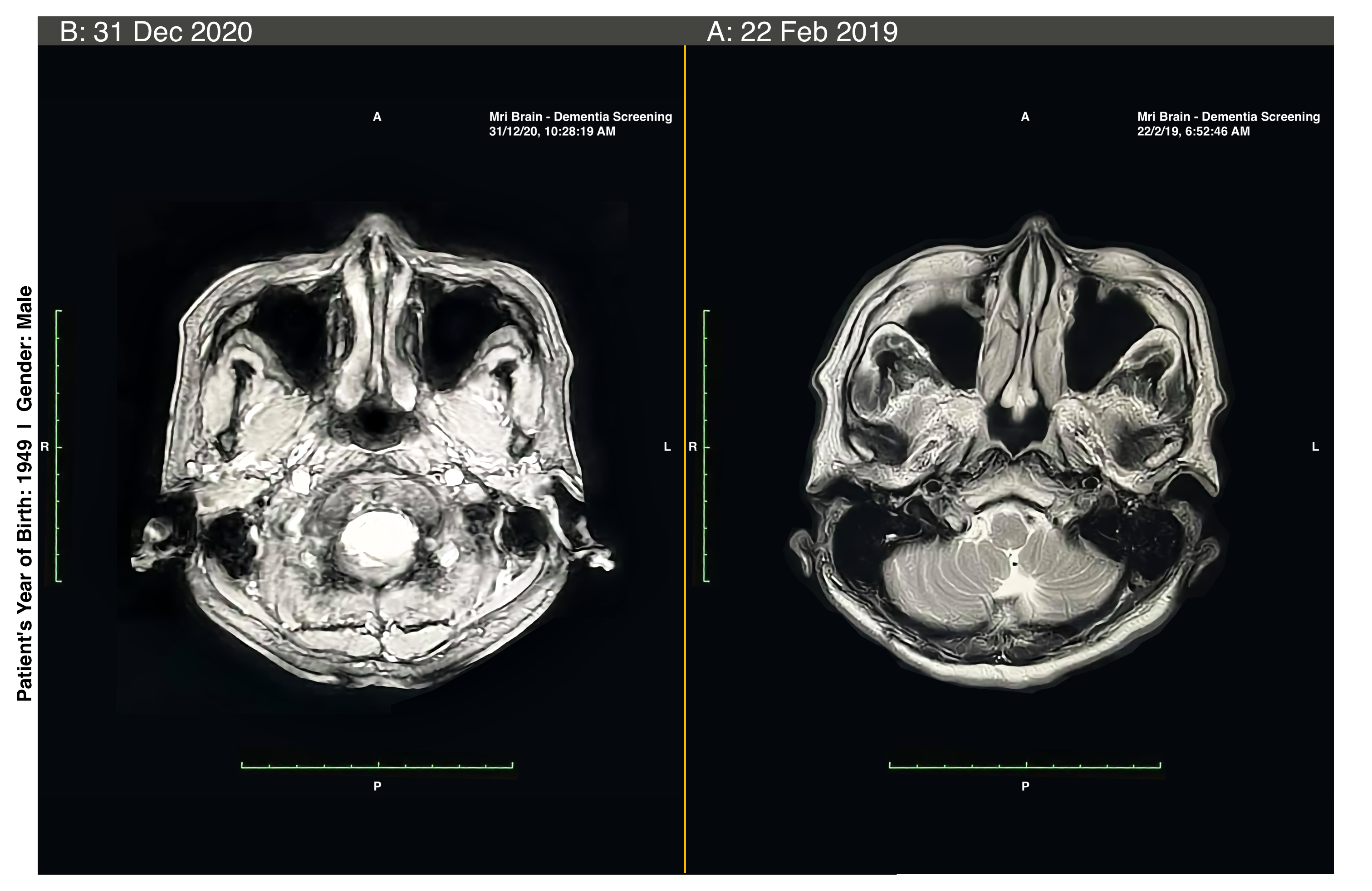
Figure 1. Sequential axial T2-weighted MRI images of the brain in a male patient (born in 1949) undergoing dementia screening.
(A) February 22, 2019: Generalized cerebral volume loss with widened extra-axial cerebrospinal fluid (CSF) spaces, prominent cortical sulci, and increased distance between the cortical surface and the inner table of the skull. The cerebellum appears relatively reduced in volume, with enlarged pericerebellar CSF spaces.
(B) December 31, 2020, following five sessions of the HEAL Method™ cranial intervention administered between September and November 2020: Apparent reduction in extra-axial CSF spaces, less prominent cortical sulci, and closer apposition of the brain parenchyma to the calvarium. The cerebellum more fully occupies the posterior fossa, with decreased surrounding CSF volume.
5. Discussion
The visual comparison between pre- and post-intervention MRI scans shows a notable change in intracranial configuration over a 22-month interval, temporally associated with the HEAL Method™ cranial intervention. While spontaneous reversal of atrophy is rare, potential mechanisms may include:
- Alterations in cranial bone dynamics that improve intracranial pressure homeostasis
- Enhanced CSF circulation, resulting in reduced compensatory CSF volume in extra-axial spaces
- Improved venous outflow, decreasing intracranial compliance abnormalities
6. Conclusion
This case demonstrates visually apparent MRI changes suggestive of reduced cerebral atrophy following a short course of HEAL Method™ cranial intervention. While the association is compelling, further controlled studies are needed to determine the reproducibility, underlying mechanisms, and clinical significance of these imaging changes.
7. Acknowledgments
The authors wish to thank the patient and his family for their kind cooperation and for providing verbal consent for the anonymous use of his medical information and imaging in this publication.
8. Conflicts of Interest
Dr. Daniel Foo is the founder of the HEAL Method™ and maintains an active clinical practice utilizing the system described in this report. This potential conflict has been disclosed and does not affect the objectivity of the report.